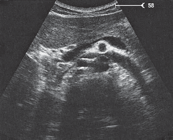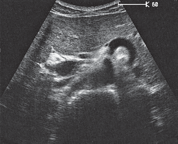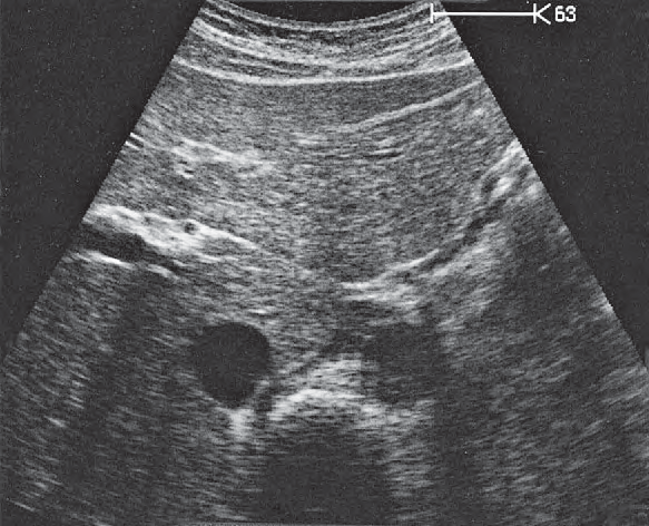
Left Renal Artery Ultrasound Anatomy
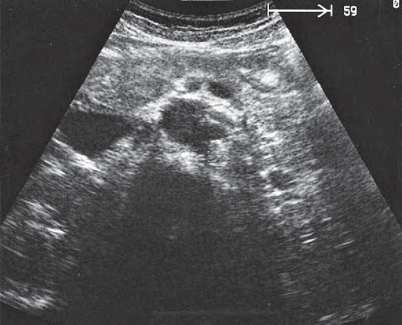
The left renal artery is a paired visceral branch of the abdominal aorta that carries oxygenated blood to the left kidney. It arises from the lateral wall of the aorta a short distance below the origin of the superior mesenteric artery, usually around the first lumbar vertebra, and runs behind the left renal vein on its way to the renal hilum.
On ultrasound the left renal artery is easiest to identify on a transverse epigastric view of the great vessels. In this plane the left renal vein crosses in front of the aorta toward the inferior vena cava, while the renal arteries leave the aorta posterolaterally. Colour and spectral Doppler confirm the vessel and show the low resistance waveform expected of a normal renal artery. Because the left renal artery also appears at the edge of many pancreatic, splenic and left kidney views, learning to recognise it in several planes makes it far easier to find on a live scan.
Left renal artery across the atlas
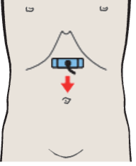
Superior mesenteric artery and splenic vein
Vessels / Aorta and Vena Cava in Suprarenal Transverse Sections, Including the Renal Vessels · Transverse plane
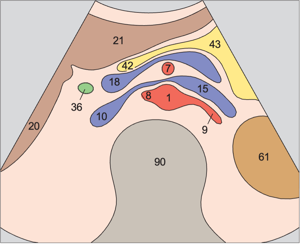
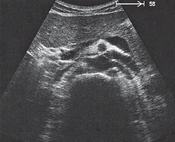
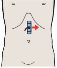
Splenic artery and vein, celiac trunk
Vessels / Splenic Artery and Vein in Longitudinal Sections · Longitudinal plane
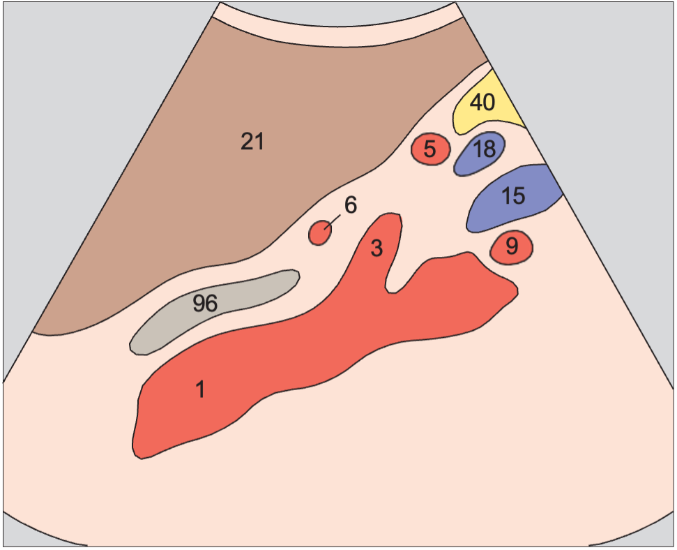
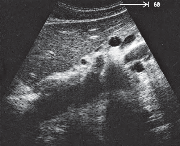
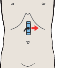
Splenic artery and vein, renal artery and vein
Vessels / Splenic Artery and Vein in Longitudinal Sections · Longitudinal plane
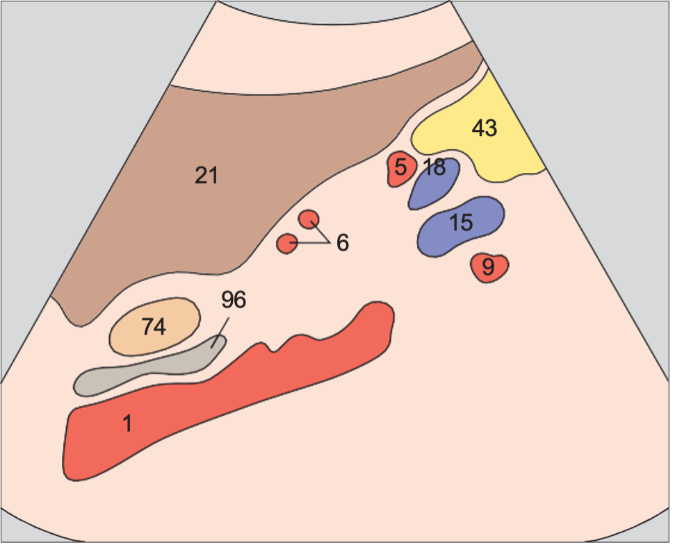
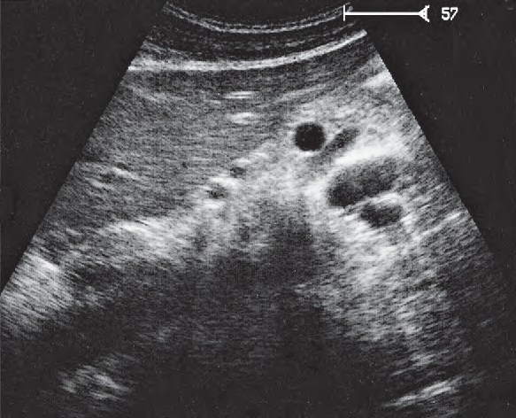
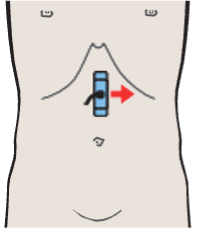
Splenic artery and vein, renal artery and vein
Vessels / Splenic Artery and Vein in Longitudinal Sections · Longitudinal plane
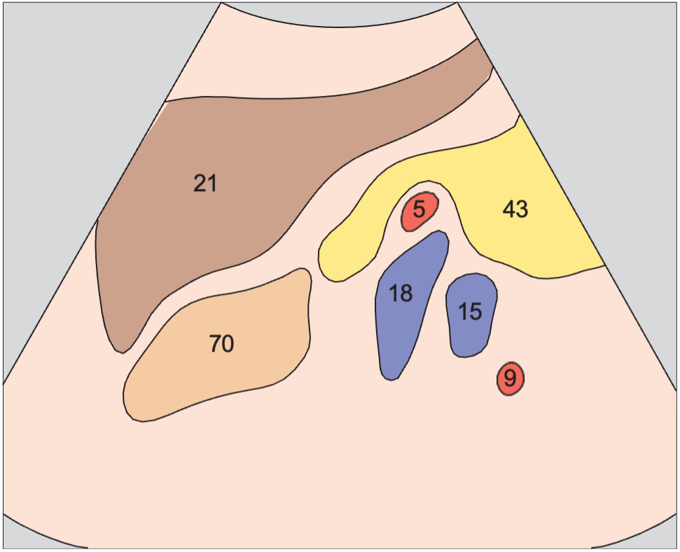
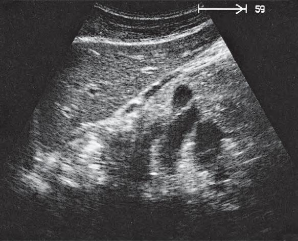
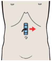
Left renal vessels, splenic artery and vein
Vessels / Left Renal Artery and Vein in Longitudinal Sections · Longitudinal plane
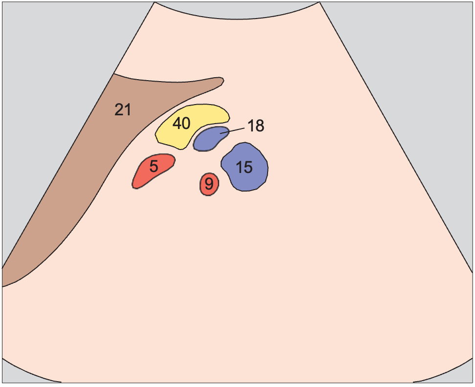
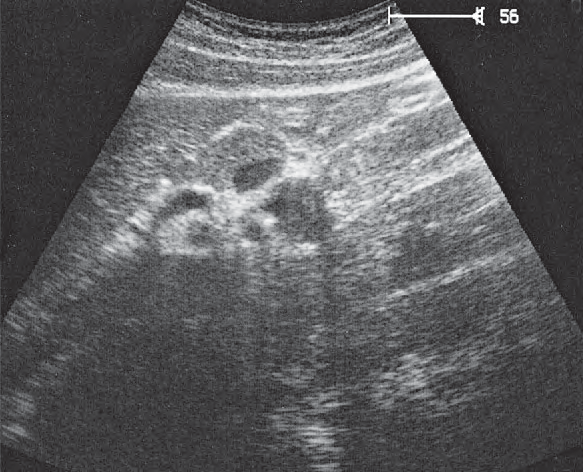
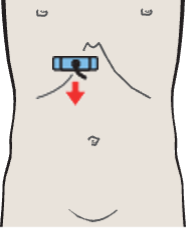
Medial and anterior hepatic segments, right and left portal vein branches
Liver / Central Portions of the Liver in Transverse Sections · Transverse plane
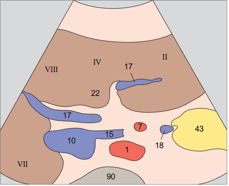
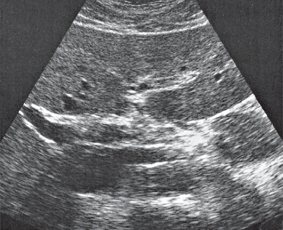
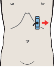
Body of pancreas, splenic vein
Pancreas / Pancreas in Longitudinal Sections · Longitudinal plane
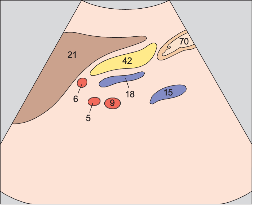
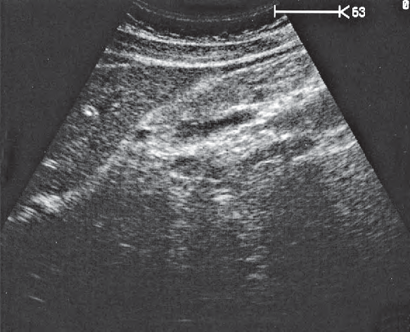
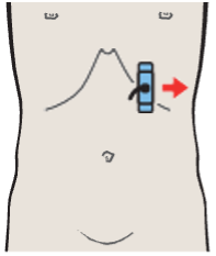
Tail of pancreas, splenic artery and vein, renal artery and vein
Pancreas / Pancreas in Longitudinal Sections · Longitudinal plane
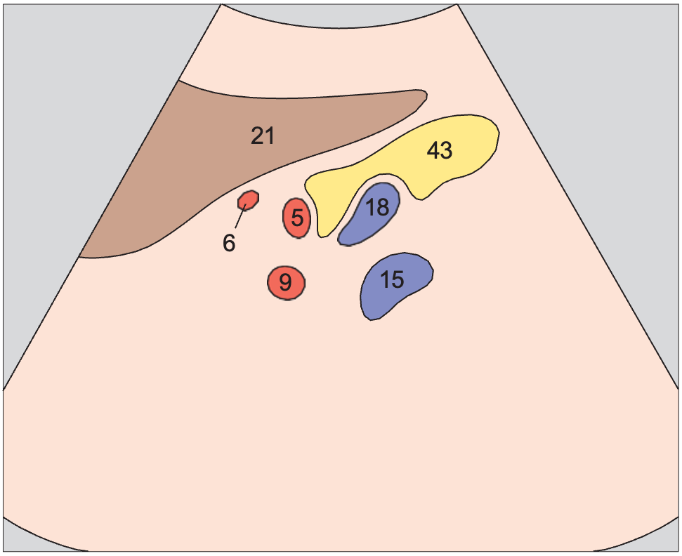
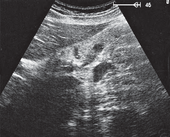
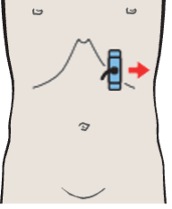
Tail of pancreas, splenic artery and vein, renal artery and vein
Pancreas / Pancreas in Longitudinal Sections · Longitudinal plane
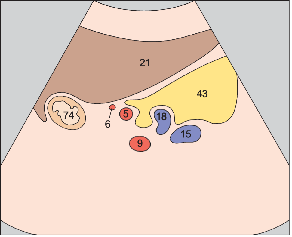
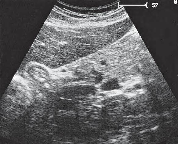
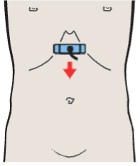
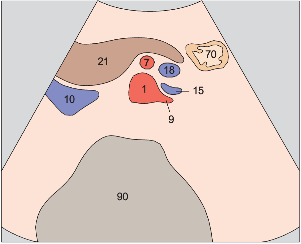
Left renal artery and vein, superior mesenteric artery and vein, aorta
Pancreas / Body of the Pancreas in Transverse Sections · Transverse plane
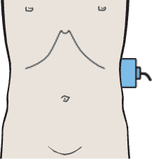
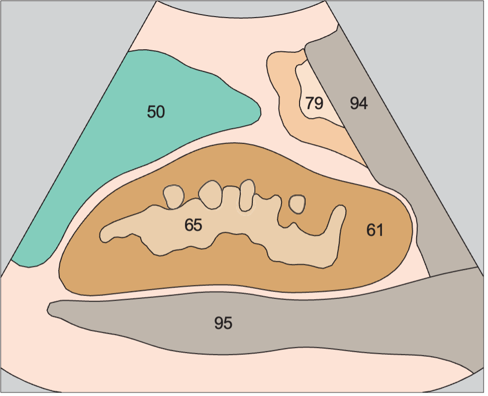
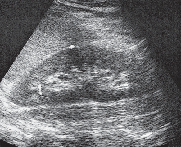
Kidney, spleen, psoas muscle
Kidneys / Longitudinal Flank Scans of the Left Kidney from Posterior to Anterior · Longitudinal plane
Anatomy diagrams, ultrasound images and probe positions are part of the Ultrasound Anatomy atlas. Colours follow the atlas legend, grouping vessels, organs and other structures by type.