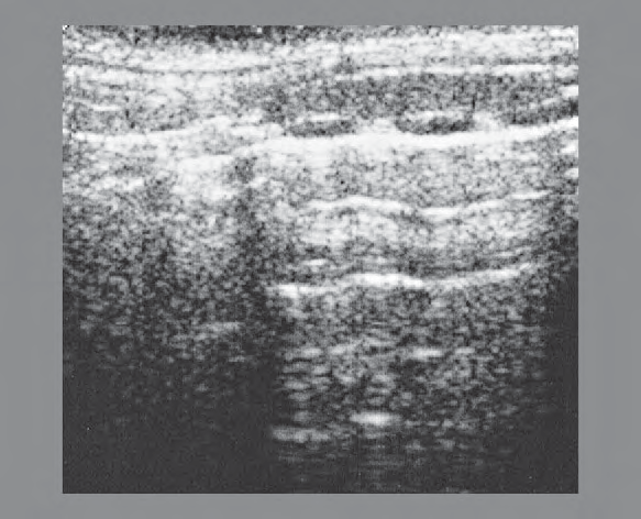
Thyroid Region
Master ultrasound anatomy with dual-view learning
Download on the App Store  Scan to install on your iPhone
Scan to install on your iPhone 244Structures
12Regions
100+Labeled Scans
Crafted by physicians, for physicians iPhone iPad coming soon Works offline Privacy-first
Features
Everything you need
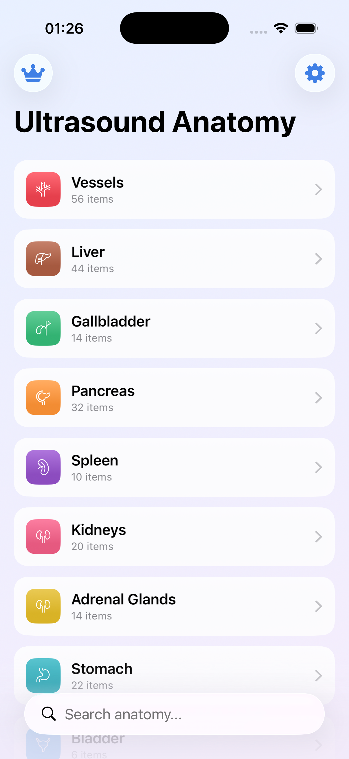
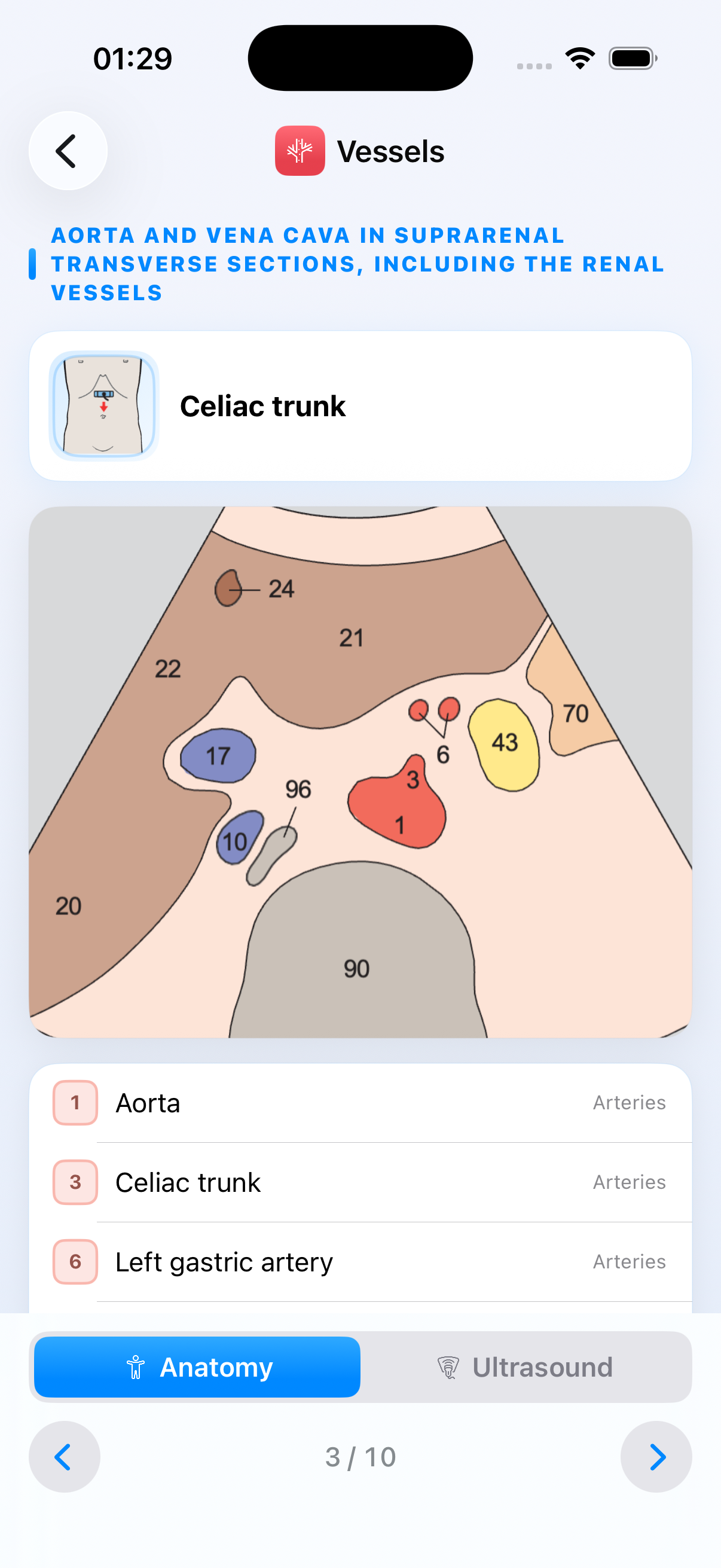
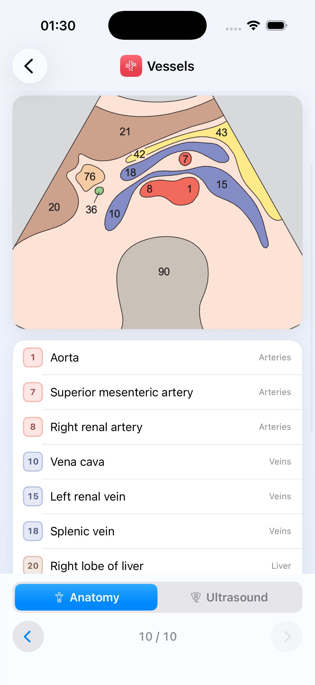
244 Anatomy Structures
Detailed diagrams for precise, exam-ready learning.
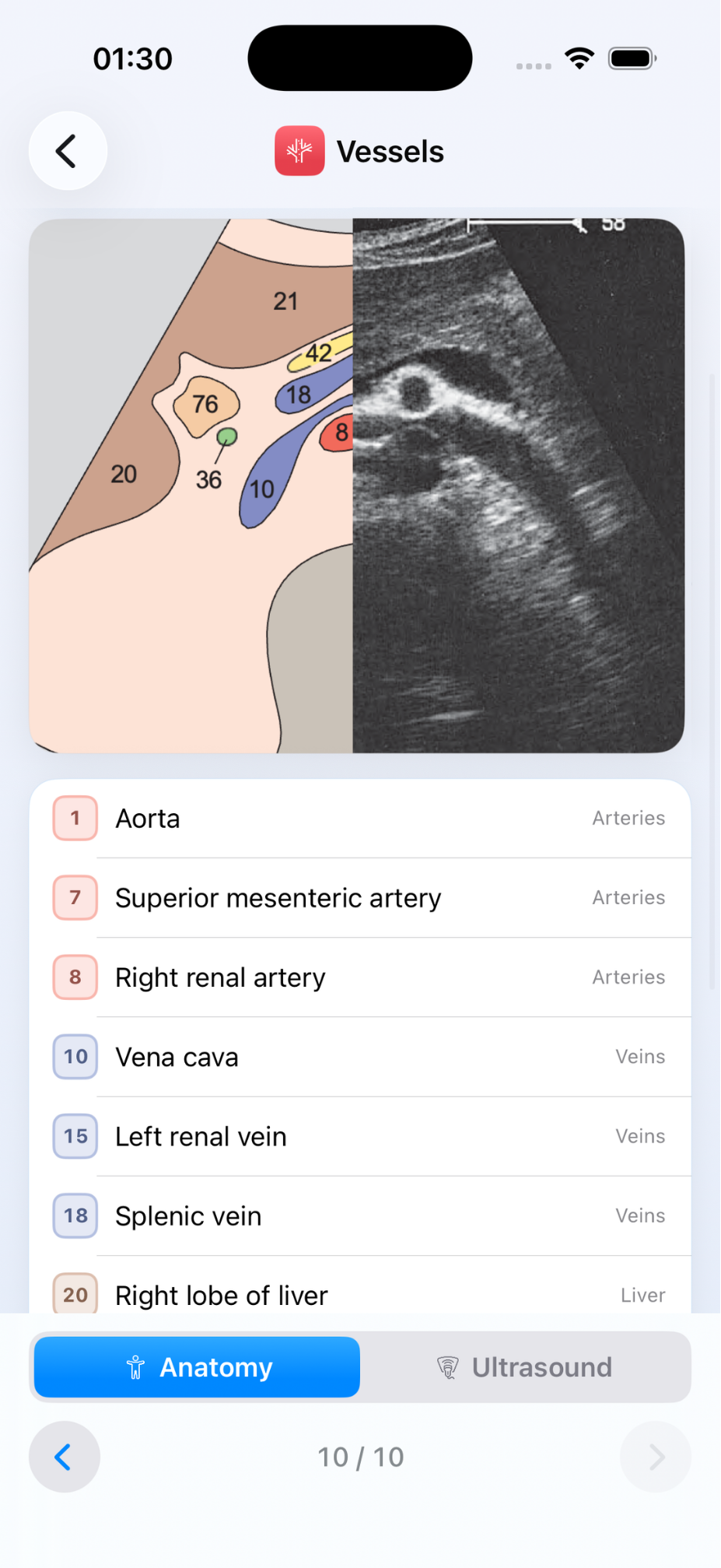
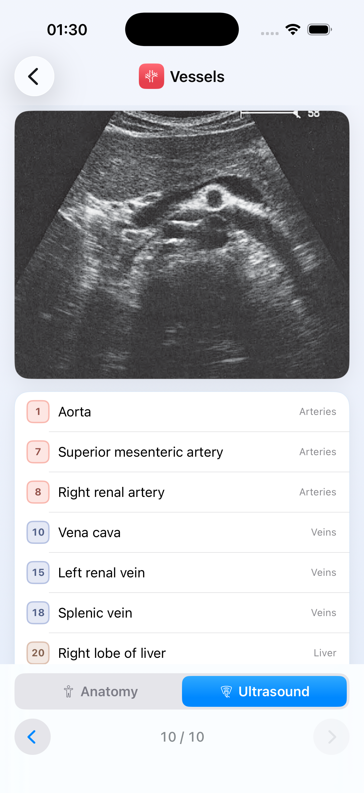
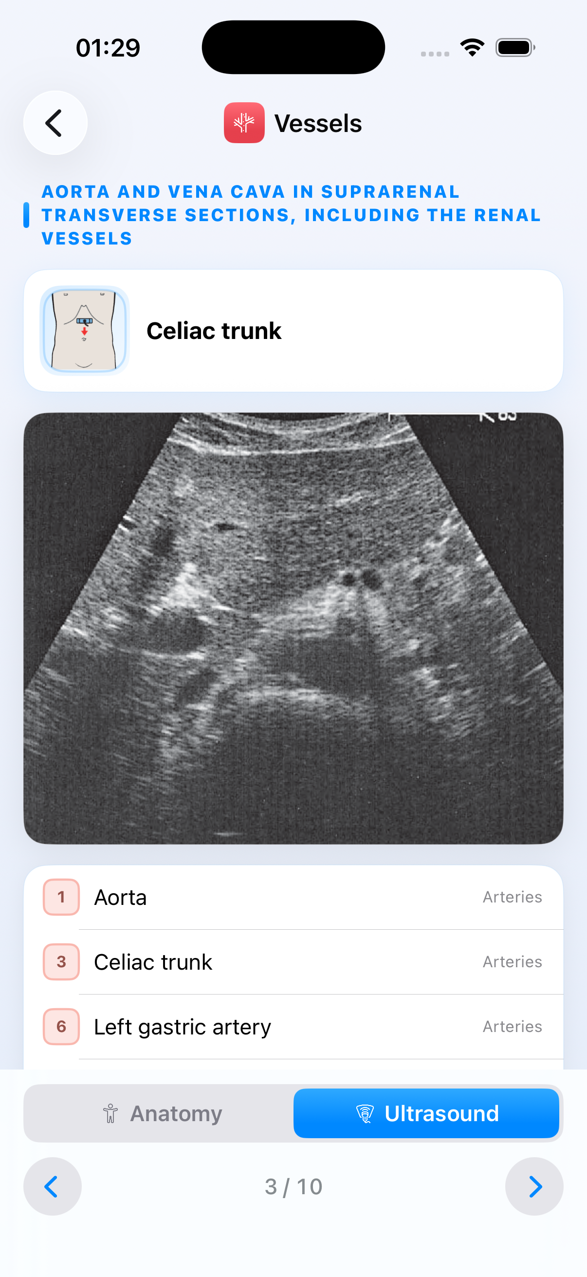
Real Ultrasound Images
Every structure paired with an actual clinical scan.
Color-Coded Legends
Arteries, veins, organs and muscles, never confused again.
Probe Placement Maps
Integrated body maps guide you to the right window.
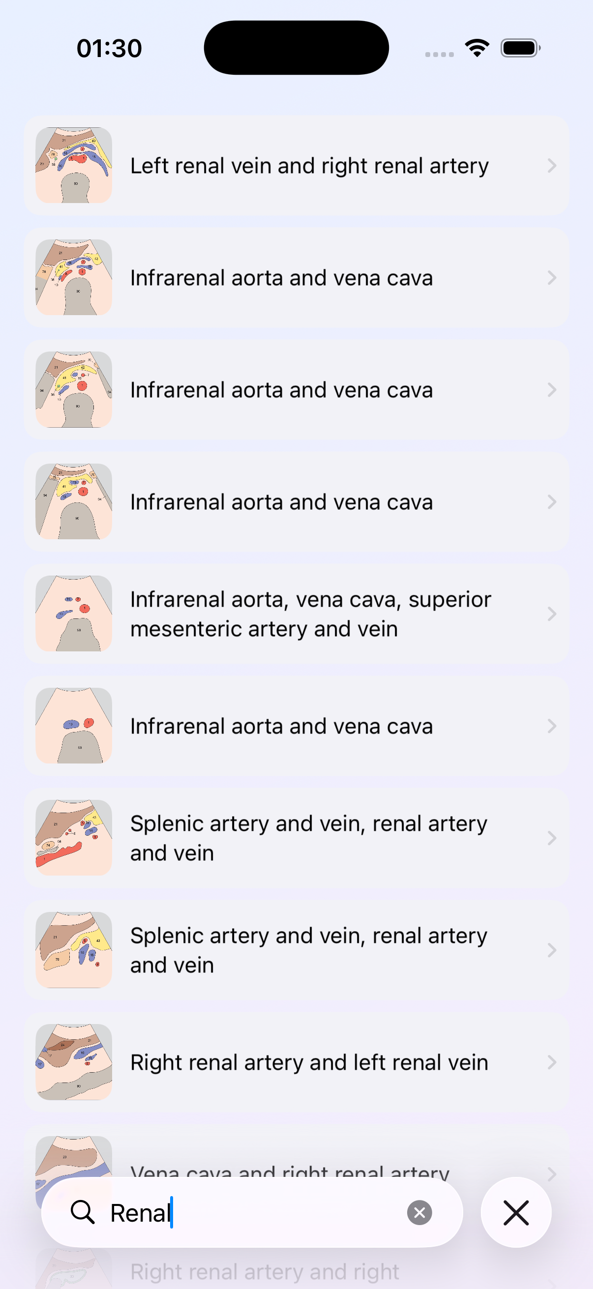
Instant Search
Find any structure or region in seconds.
12 Regions Covered
Complete anatomical coverage, head to toe.
Screenshots
See it in action
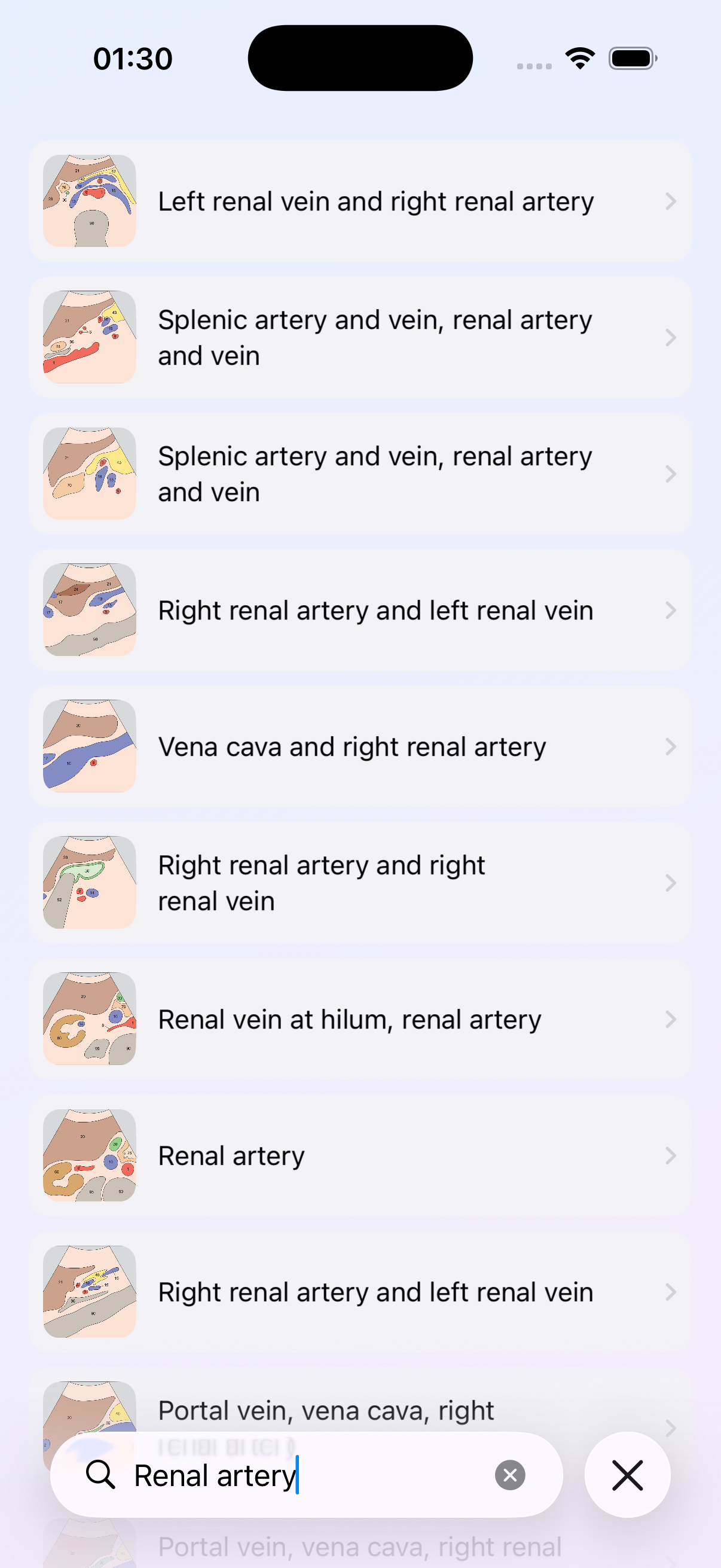
← Swipe to explore →
FAQ
Frequently asked questions
Who is this app for?
Medical and sonography students, radiologists, ER physicians, ultrasound technicians and POCUS learners — anyone who wants to master what they see on the screen.
Does it work offline?
Yes. Once installed, the full atlas of structures, diagrams and scans is available without an internet connection.
What is dual-view learning?
Switch instantly between a professional anatomy diagram and a real-world ultrasound image of the same region to bridge theory and practice.
How much does it cost?
The app is free to download and includes a subscription that unlocks the complete atlas. See the App Store for current pricing.
From the blog
Learn more, scan better
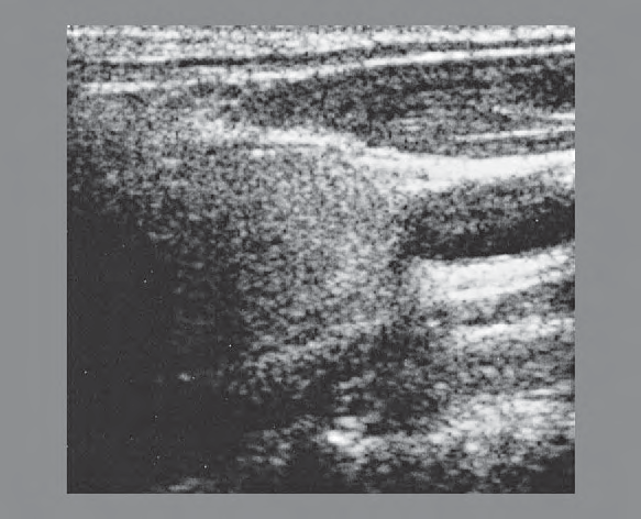
Thyroid Region
Common Carotid Artery Ultrasound Anatomy
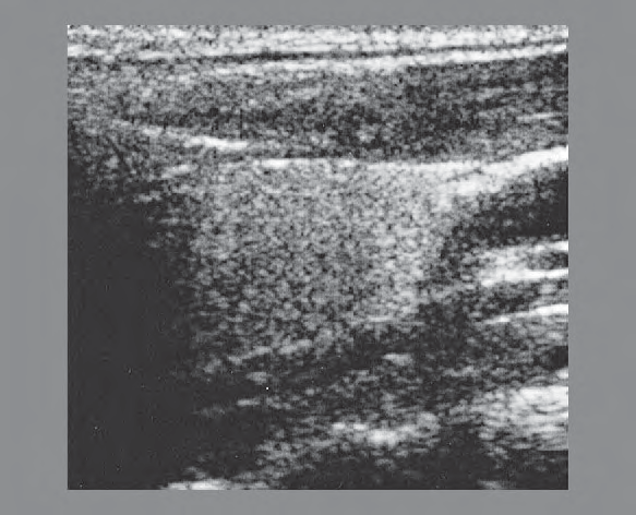
Thyroid Region
Internal Jugular Vein Ultrasound Anatomy

Get started
Ready to master it? Download Ultrasound Anatomy.
Free on the App Store. Scan the code or tap to install on your iPhone.