Ultrasound Anatomy

Acoustic Shadow Ultrasound Anatomy
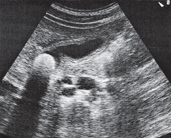
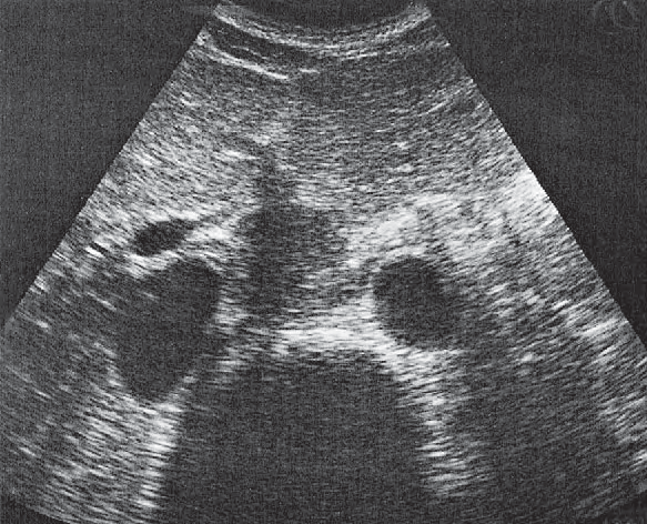
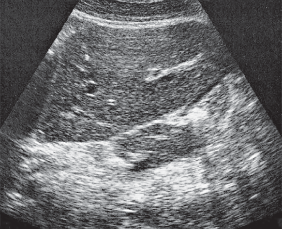
Acoustic shadow ultrasound image

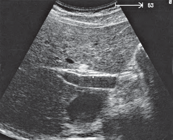
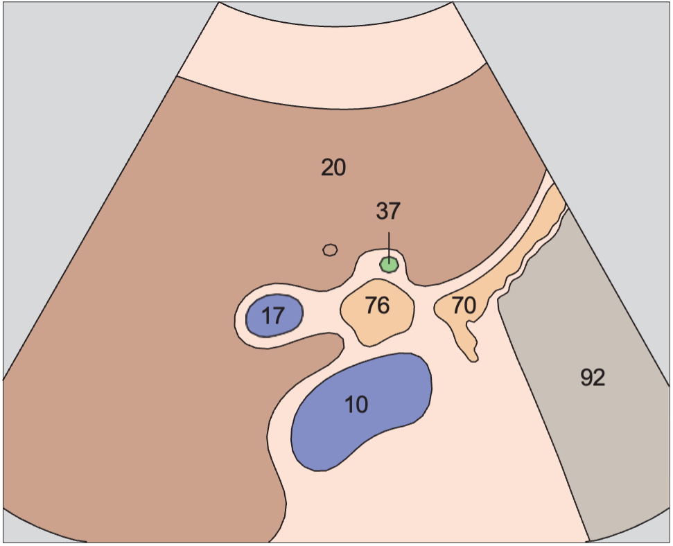
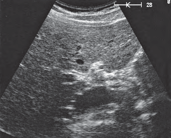
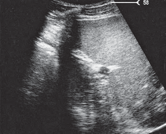
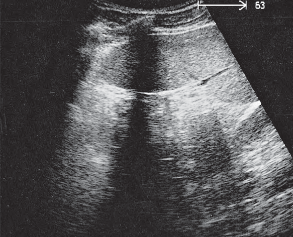
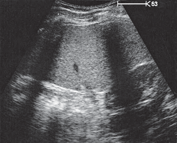
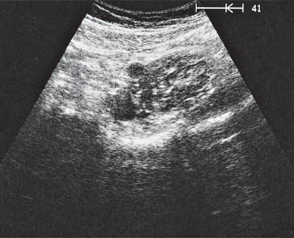
Acoustic shadow ultrasound anatomy is presented here as a labeled cross-sectional atlas. Every view pairs a color-coded anatomy diagram with the matching real ultrasound image, so you can learn the expected shape and position of the acoustic shadow and then recognise it on a real scan.

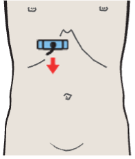
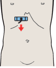
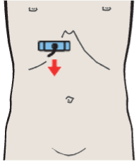
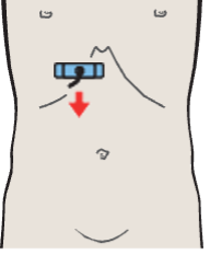
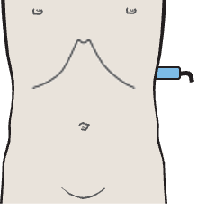
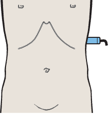
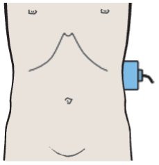
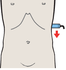
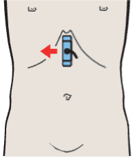
In the atlas legend the acoustic shadow belongs to the general structures group and is shown in a consistent colour wherever it appears. The small body marker on each card shows where the transducer sits for that view, which makes it easier to reproduce the image at the bedside.

Acoustic shadow appears in 14 labeled views in this atlas. Each card below shows the anatomy diagram, the real ultrasound image and the probe position for that scan. Coloured dots list every structure labeled in the view, with acoustic shadow highlighted.

14Scan views

Acoustic shadow across the atlas

Probe position, Right renal artery and right renal vein

Right renal artery and right renal vein

Vessels / Renal Artery and Vein in Longitudinal Sections · Longitudinal plane
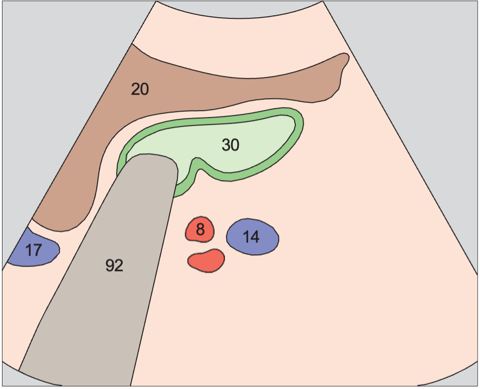
AnatomyAcoustic shadow anatomy diagram, Right renal artery and right renal vein
UltrasoundAcoustic shadow ultrasound image, Right renal artery and right renal vein
8Right renal artery14Right renal vein17Portal vein20Right lobe of liver30Gallbladder92Acoustic shadow
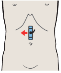
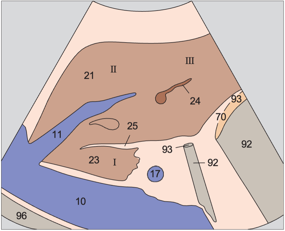
Probe position, Left lobe of liver, ligamentum teres, boundary between lateral and medial segments, caudate lobe

Left lobe of liver, ligamentum teres, boundary between lateral and medial segments, caudate lobe

Liver / Liver in Longitudinal Sections · Longitudinal plane
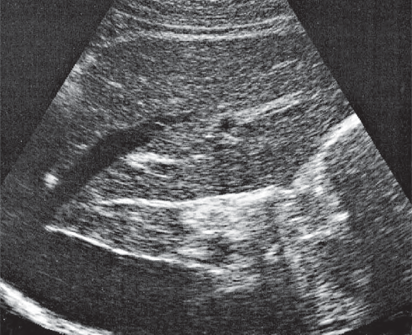
AnatomyAcoustic shadow anatomy diagram, Left lobe of liver, ligamentum teres, boundary between lateral and medial segments, caudate lobe
UltrasoundAcoustic shadow ultrasound image, Left lobe of liver, ligamentum teres, boundary between lateral and medial segments, caudate lobe
10Vena cava11Left hepatic vein17Portal vein21Left lobe of liver23Caudate lobe24Ligamentum teres25Ligamentum venosum70Stomach92Acoustic shadow93Gas96Diaphragm
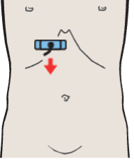
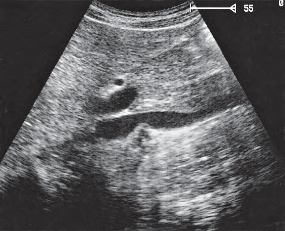
Probe position, Right portal vein branch, ligamentum venosum

Right portal vein branch, ligamentum venosum

Gallbladder / Gallbladder in Upper Abdominal Transverse Sections · Transverse plane

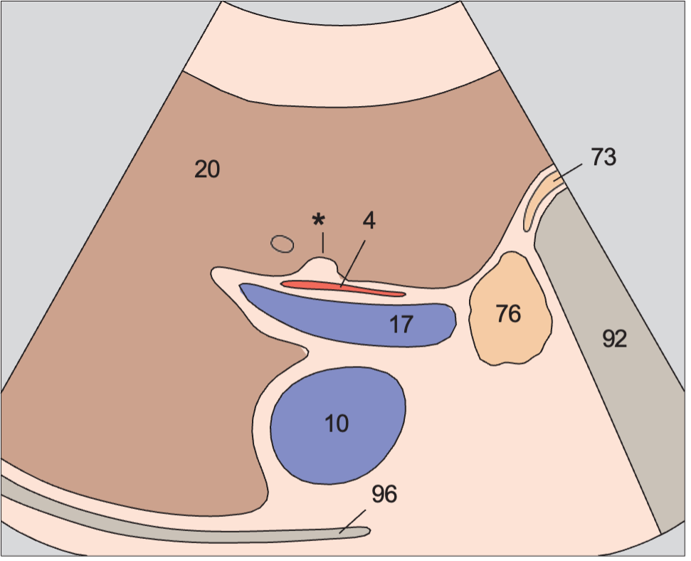
AnatomyAcoustic shadow anatomy diagram, Right portal vein branch, ligamentum venosum
UltrasoundAcoustic shadow ultrasound image, Right portal vein branch, ligamentum venosum
4Hepatic artery10Vena cava17Portal vein20Right lobe of liver73Antrum of stomach76Duodenum92Acoustic shadow96Diaphragm
Probe position, Gallbladder neck

Gallbladder neck

Gallbladder / Gallbladder in Upper Abdominal Transverse Sections · Transverse plane

AnatomyAcoustic shadow anatomy diagram, Gallbladder neck
UltrasoundAcoustic shadow ultrasound image, Gallbladder neck
10Vena cava17Portal vein20Right lobe of liver37Cystic duct70Stomach76Duodenum92Acoustic shadow
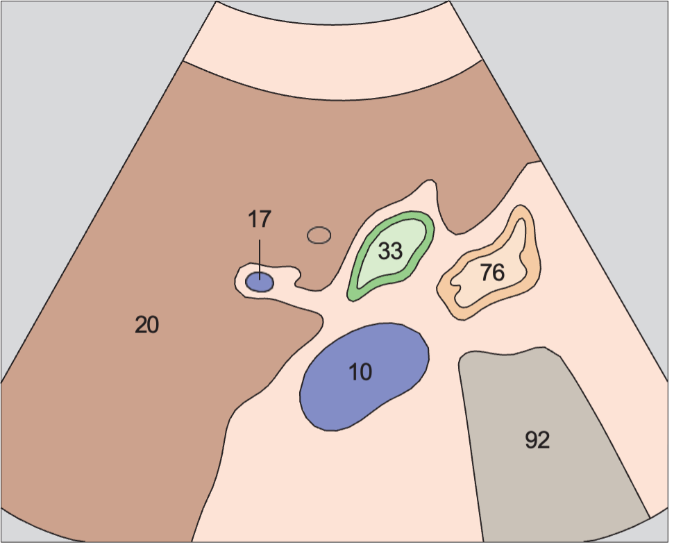
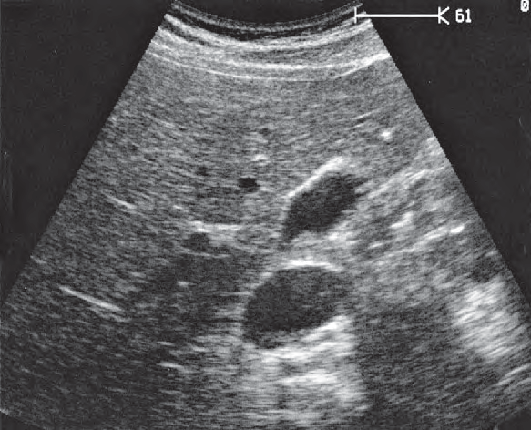
Probe position, Junction of neck and body of gallbladder

Junction of neck and body of gallbladder

Gallbladder / Gallbladder in Upper Abdominal Transverse Sections · Transverse plane

AnatomyAcoustic shadow anatomy diagram, Junction of neck and body of gallbladder
UltrasoundAcoustic shadow ultrasound image, Junction of neck and body of gallbladder
10Vena cava17Portal vein20Right lobe of liver33Neck of gallbladder76Duodenum92Acoustic shadow
Probe position, Body of gallbladder

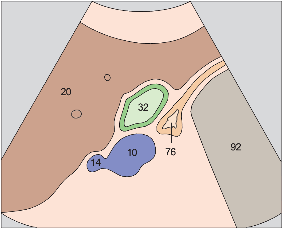
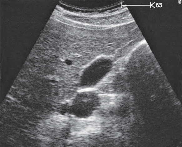
Body of gallbladder

Gallbladder / Gallbladder in Upper Abdominal Transverse Sections · Transverse plane

AnatomyAcoustic shadow anatomy diagram, Body of gallbladder
UltrasoundAcoustic shadow ultrasound image, Body of gallbladder
10Vena cava14Right renal vein20Right lobe of liver32Body of gallbladder76Duodenum92Acoustic shadow
Probe position, Gallbladder fundus

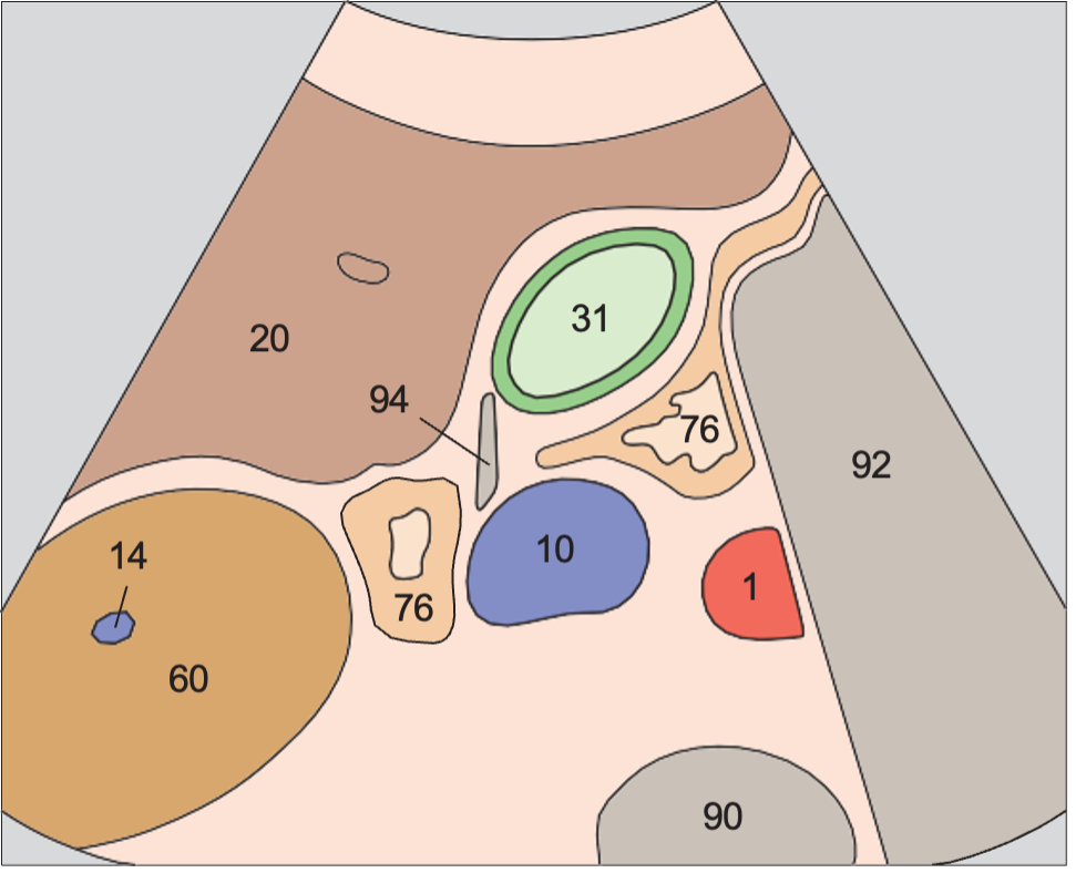
Gallbladder fundus

Gallbladder / Gallbladder in Upper Abdominal Transverse Sections · Transverse plane

AnatomyAcoustic shadow anatomy diagram, Gallbladder fundus
UltrasoundAcoustic shadow ultrasound image, Gallbladder fundus
1Aorta10Vena cava14Right renal vein20Right lobe of liver31Fundus of gallbladder60Right kidney76Duodenum90Spinal column92Acoustic shadow94Artifact
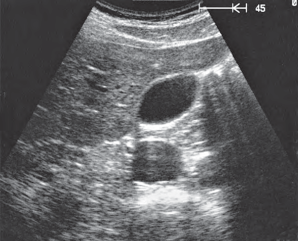
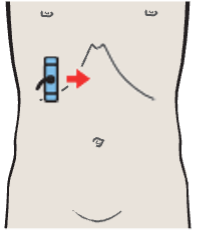
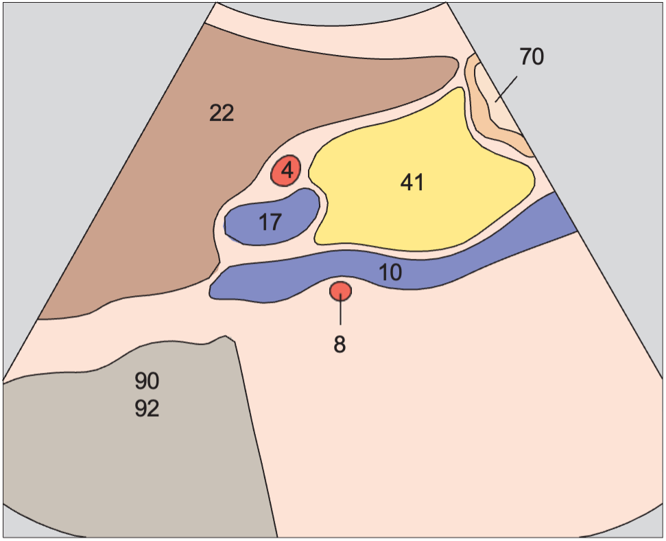
Probe position, Head of pancreas, hilar vessels, vena cava

Head of pancreas, hilar vessels, vena cava

Pancreas / Pancreas in Longitudinal Sections · Longitudinal plane
AnatomyAcoustic shadow anatomy diagram, Head of pancreas, hilar vessels, vena cava
UltrasoundAcoustic shadow ultrasound image, Head of pancreas, hilar vessels, vena cava
4Hepatic artery8Right renal artery10Vena cava17Portal vein22Quadrate lobe41Head of pancreas70Stomach90Spinal column92Acoustic shadow
Probe position, Spleen, kidney, stomach

Spleen, kidney, stomach

Spleen / Transverse Flank Scans of the Spleen · Transverse plane
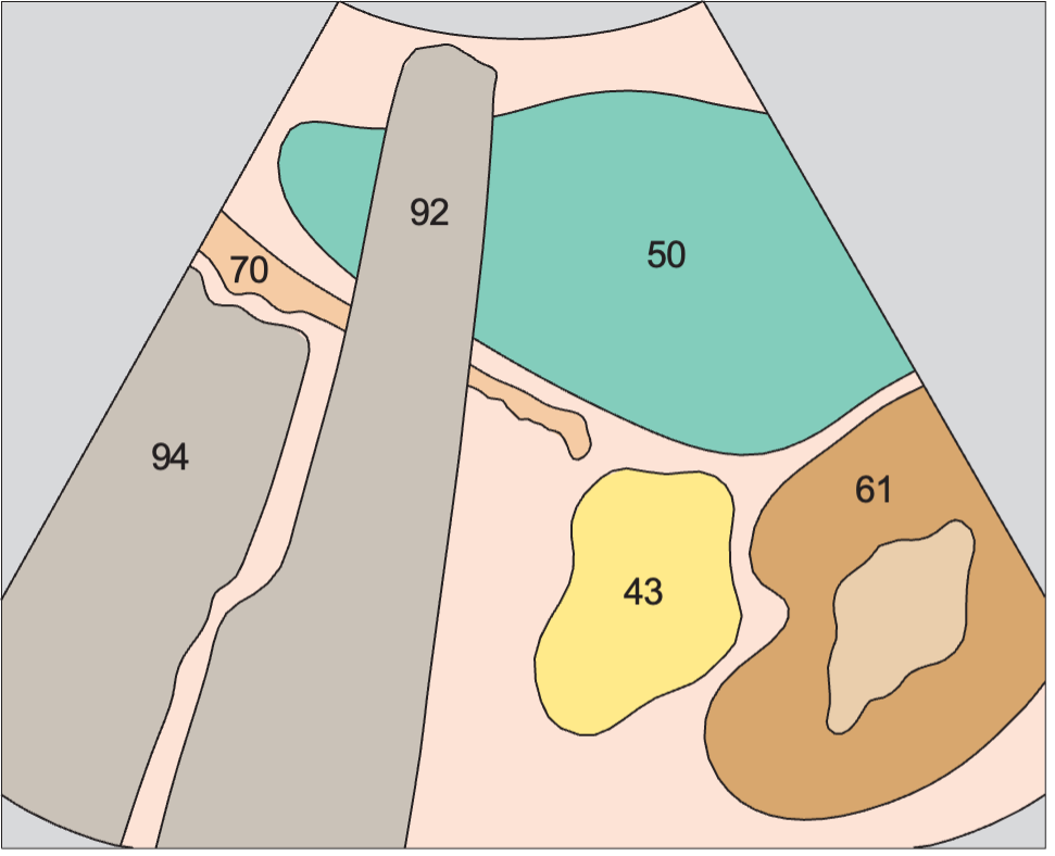
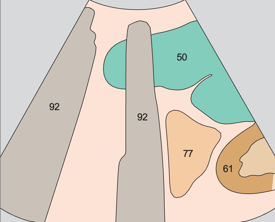
AnatomyAcoustic shadow anatomy diagram, Spleen, kidney, stomach
UltrasoundAcoustic shadow ultrasound image, Spleen, kidney, stomach
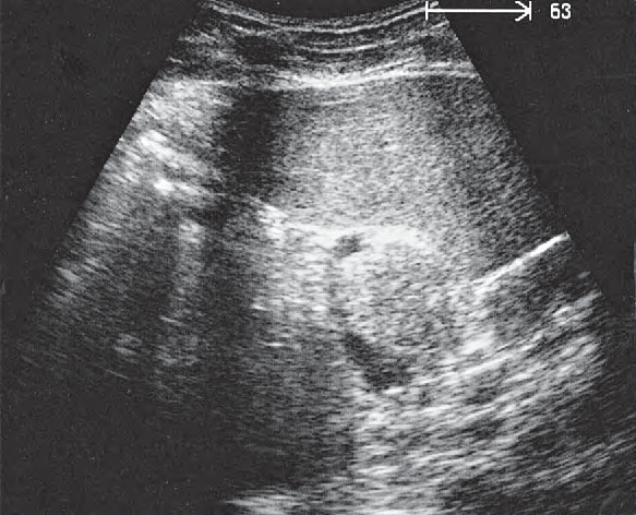
43Tail of pancreas50Spleen61Left kidney70Stomach92Acoustic shadow94Artifact
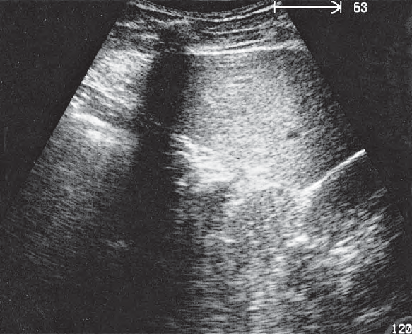
Probe position, Spleen, kidney, pancreas

Spleen, kidney, pancreas

Spleen / Transverse Flank Scans of the Spleen · Transverse plane
AnatomyAcoustic shadow anatomy diagram, Spleen, kidney, pancreas
UltrasoundAcoustic shadow ultrasound image, Spleen, kidney, pancreas
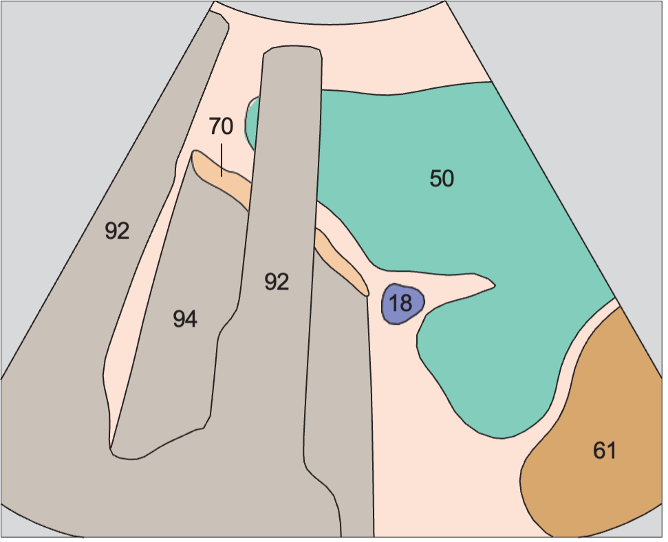
18Splenic vein43Tail of pancreas50Spleen61Left kidney70Stomach92Acoustic shadow94Artifact
Probe position, Spleen, stomach

Spleen, stomach

Spleen / Transverse Flank Scans of the Spleen · Transverse plane
AnatomyAcoustic shadow anatomy diagram, Spleen, stomach
UltrasoundAcoustic shadow ultrasound image, Spleen, stomach
18Splenic vein50Spleen61Left kidney70Stomach92Acoustic shadow94Artifact
Probe position, Spleen, small bowel

Spleen, small bowel

Spleen / Transverse Flank Scans of the Spleen · Transverse plane
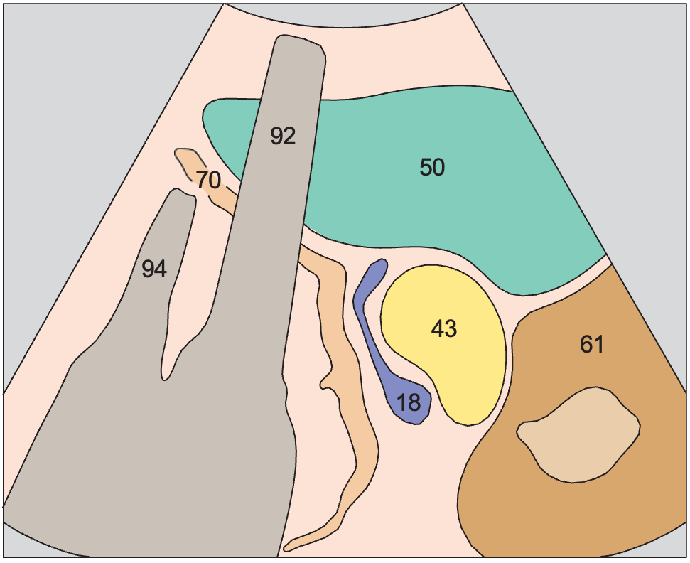
AnatomyAcoustic shadow anatomy diagram, Spleen, small bowel
UltrasoundAcoustic shadow ultrasound image, Spleen, small bowel
50Spleen61Left kidney77Small bowel92Acoustic shadow
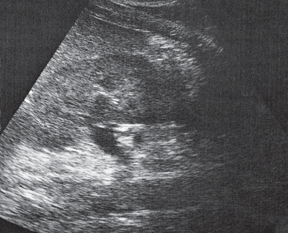
Probe position, Kidney, renal vein, spleen, aorta

Kidney, renal vein, spleen, aorta

Kidneys / Longitudinal Flank Scans of the Left Kidney from Posterior to Anterior · Longitudinal plane
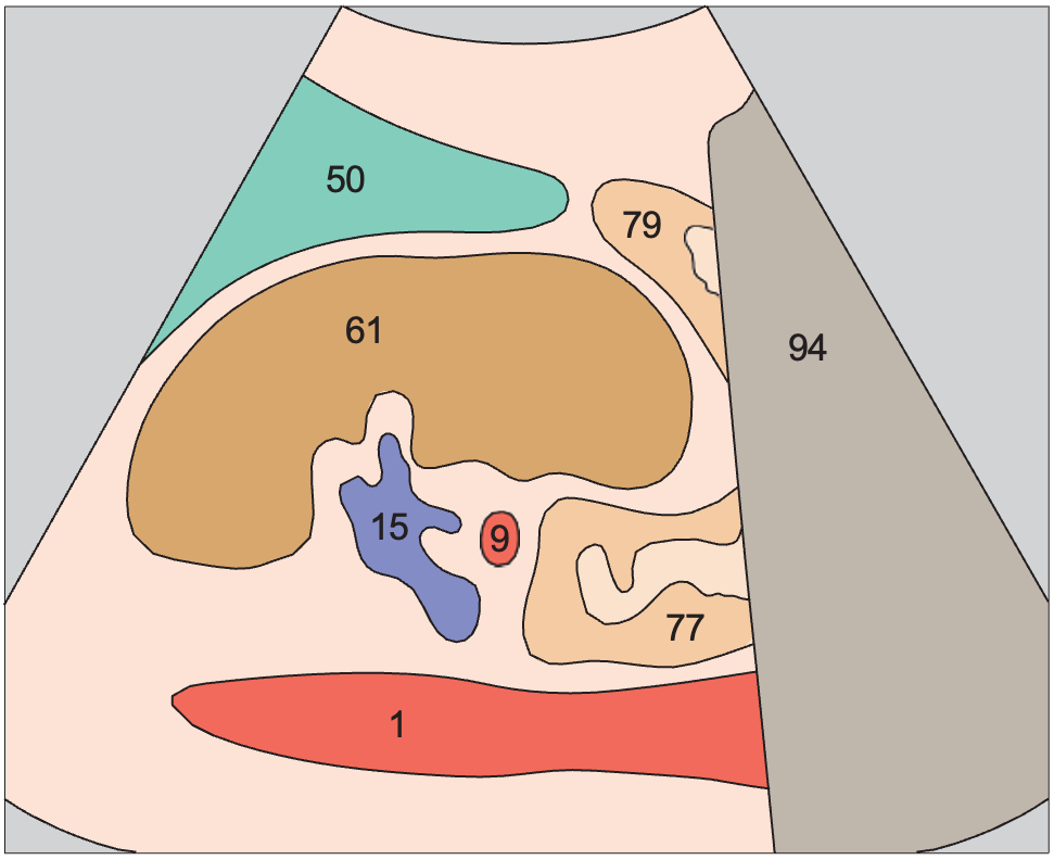
AnatomyAcoustic shadow anatomy diagram, Kidney, renal vein, spleen, aorta
UltrasoundAcoustic shadow ultrasound image, Kidney, renal vein, spleen, aorta
1Aorta15Left renal vein50Spleen61Left kidney77Small bowel92Acoustic shadow
Probe position, Kidney, spleen, bowel

Kidney, spleen, bowel

Kidneys / Transverse Flank Scans of the Left Kidney from Above Downward · Transverse plane
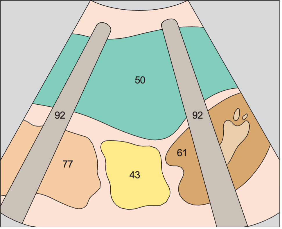
AnatomyAcoustic shadow anatomy diagram, Kidney, spleen, bowel
UltrasoundAcoustic shadow ultrasound image, Kidney, spleen, bowel
15Left renal vein50Spleen61Left kidney90Spinal column92Acoustic shadow95Psoas muscle

Anatomy diagrams, ultrasound images and probe positions are part of the Ultrasound Anatomy atlas. Colours follow the atlas legend, grouping vessels, organs and other structures by type.

Ultrasound Anatomy

Take Ultrasound Anatomy with you

QR code to download
Share

Leave a comment

Your email address will not be published. Required fields are marked *

Secret Link