Ultrasound Anatomy

Iliac Vein Ultrasound Anatomy
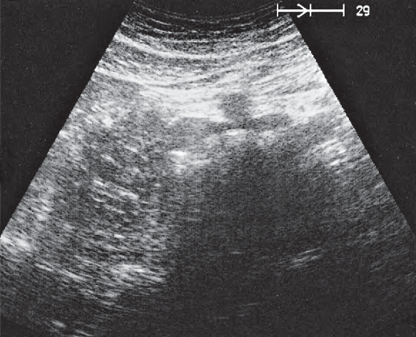
Iliac vein ultrasound image

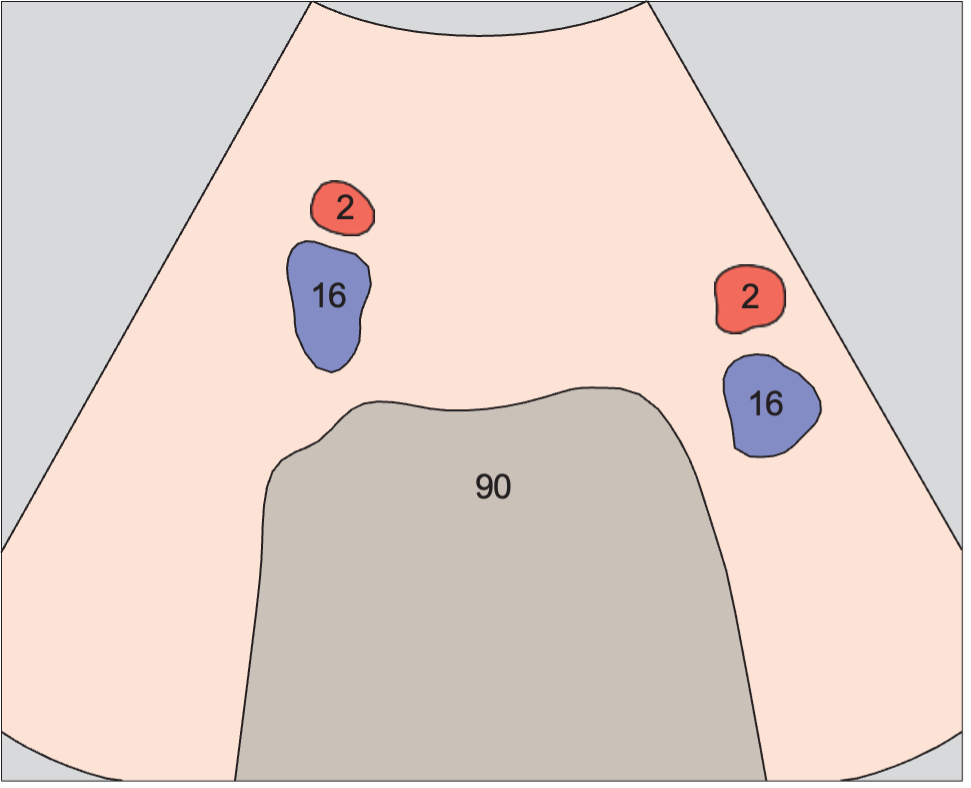
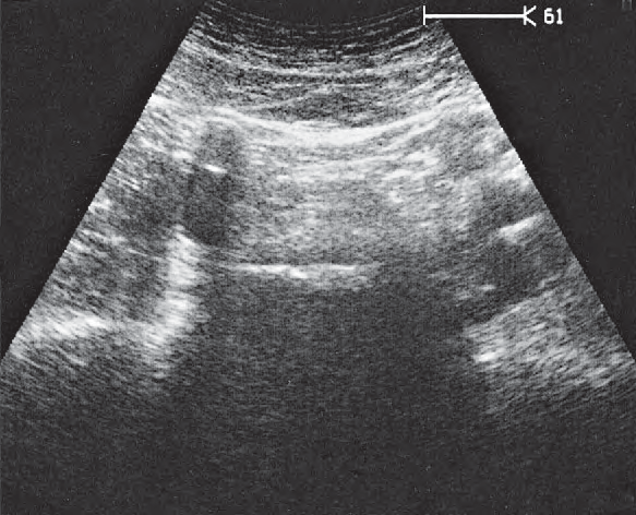
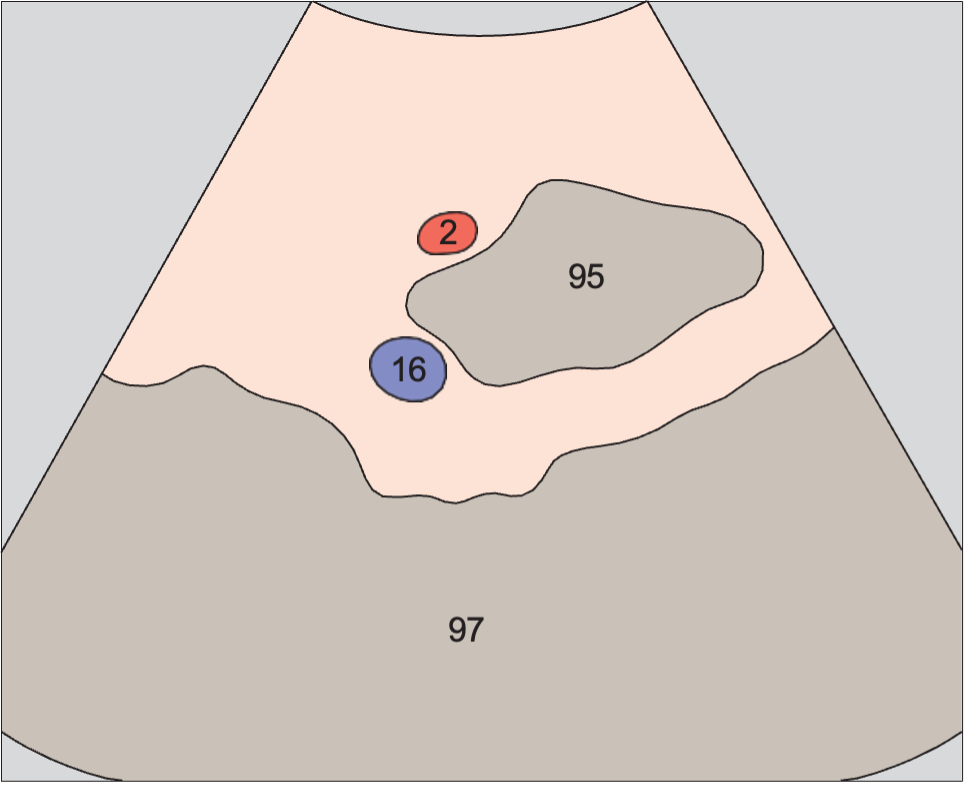
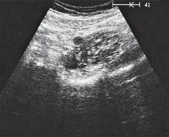
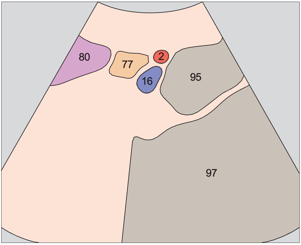
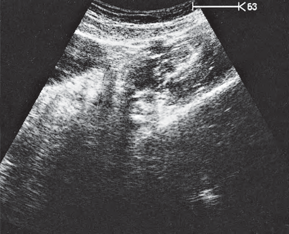
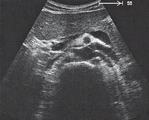
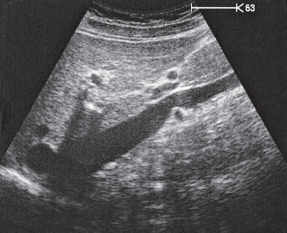
Iliac vein ultrasound anatomy is presented here as a labeled cross-sectional atlas. Every view pairs a color-coded anatomy diagram with the matching real ultrasound image, so you can learn the expected shape and position of the iliac vein and then recognise it on a real scan.

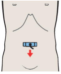
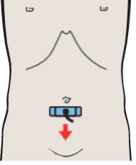
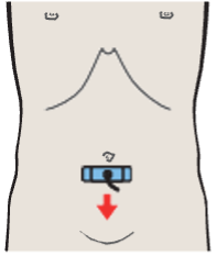
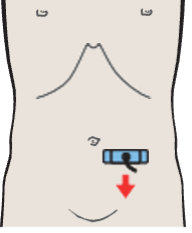
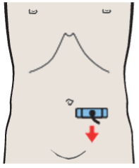
In the atlas legend the iliac vein belongs to the veins group and is shown in a consistent colour wherever it appears. The small body marker on each card shows where the transducer sits for that view, which makes it easier to reproduce the image at the bedside.

Iliac vein appears in 7 labeled views in this atlas. Each card below shows the anatomy diagram, the real ultrasound image and the probe position for that scan. Coloured dots list every structure labeled in the view, with iliac vein highlighted.

7Scan views

Iliac vein across the atlas

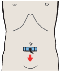
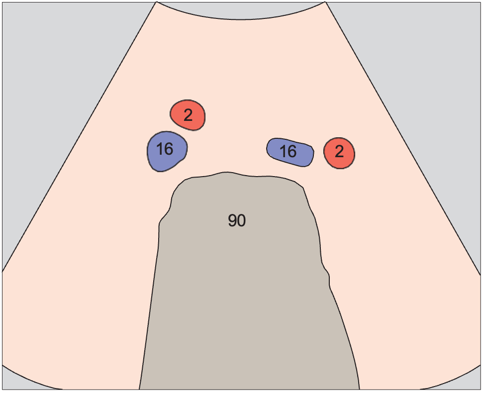
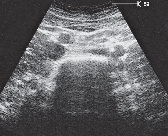
Probe position, Confluence of iliac veins

Confluence of iliac veins

Vessels / Aorta and Vena Cava in Infrarenal Transverse Sections · Transverse plane
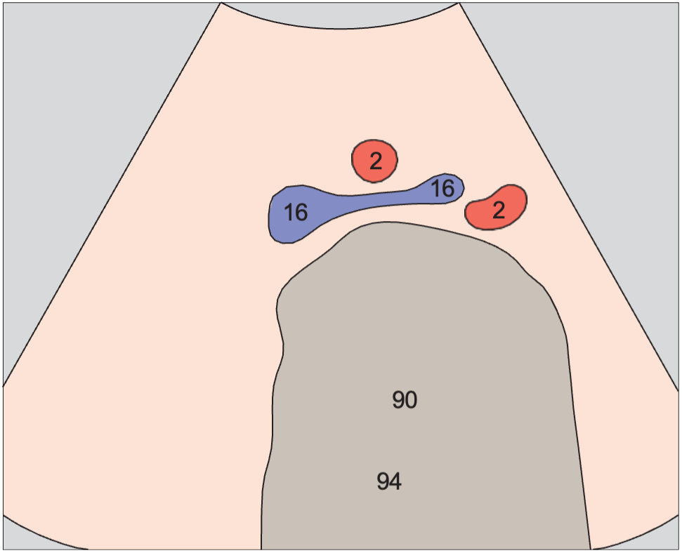
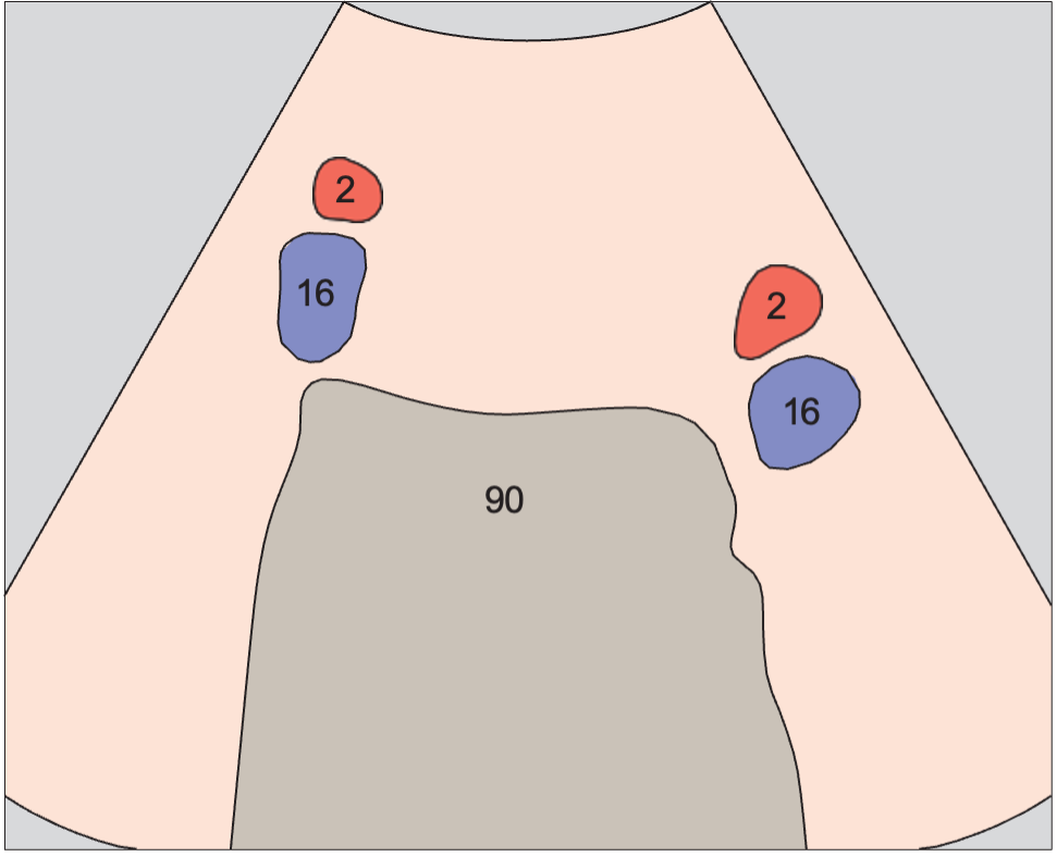
AnatomyIliac vein anatomy diagram, Confluence of iliac veins
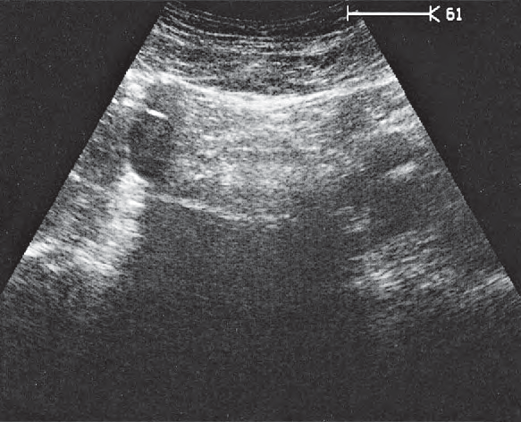
UltrasoundIliac vein ultrasound image, Confluence of iliac veins
2Iliac artery16Iliac vein90Spinal column94Artifact
Probe position, Iliac vessels

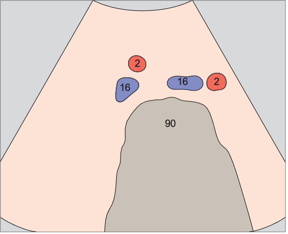
Iliac vessels

Vessels / Aorta and Vena Cava in Infrarenal Transverse Sections · Transverse plane
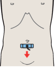
AnatomyIliac vein anatomy diagram, Iliac vessels
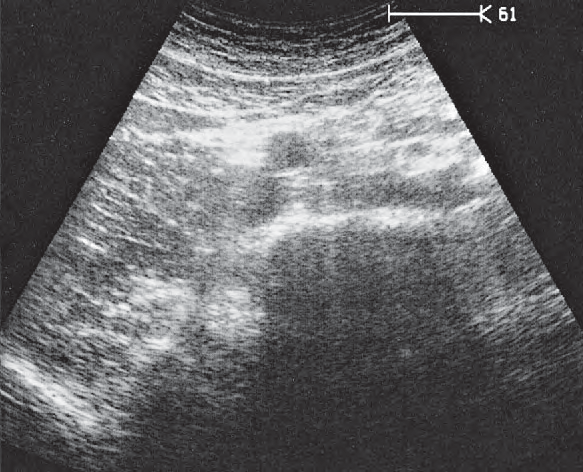
UltrasoundIliac vein ultrasound image, Iliac vessels
2Iliac artery16Iliac vein90Spinal column
Probe position, Iliac vessels

Iliac vessels

Vessels / Aorta and Vena Cava in Infrarenal Transverse Sections · Transverse plane
AnatomyIliac vein anatomy diagram, Iliac vessels
UltrasoundIliac vein ultrasound image, Iliac vessels
2Iliac artery16Iliac vein90Spinal column
Probe position, Iliac vessels

Iliac vessels

Vessels / Aorta and Vena Cava in Infrarenal Transverse Sections · Transverse plane
AnatomyIliac vein anatomy diagram, Iliac vessels
UltrasoundIliac vein ultrasound image, Iliac vessels
2Iliac artery16Iliac vein90Spinal column
Probe position, Iliac vessels

Iliac vessels

Vessels / Aorta and Vena Cava in Infrarenal Transverse Sections · Transverse plane
AnatomyIliac vein anatomy diagram, Iliac vessels
UltrasoundIliac vein ultrasound image, Iliac vessels
2Iliac artery16Iliac vein90Spinal column
Probe position, Left iliac vessels

Left iliac vessels

Vessels / Aorta and Vena Cava in Infrarenal Transverse Sections · Transverse plane
AnatomyIliac vein anatomy diagram, Left iliac vessels
UltrasoundIliac vein ultrasound image, Left iliac vessels
2Iliac artery16Iliac vein95Psoas muscle97Pelvic bone
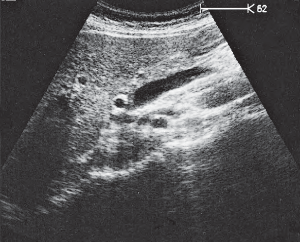
Probe position, Left iliac vessels

Left iliac vessels

Vessels / Aorta and Vena Cava in Infrarenal Transverse Sections · Transverse plane
AnatomyIliac vein anatomy diagram, Left iliac vessels
UltrasoundIliac vein ultrasound image, Left iliac vessels
2Iliac artery16Iliac vein77Small bowel80Bladder95Psoas muscle97Pelvic bone

Anatomy diagrams, ultrasound images and probe positions are part of the Ultrasound Anatomy atlas. Colours follow the atlas legend, grouping vessels, organs and other structures by type.

Ultrasound Anatomy

Take Ultrasound Anatomy with you

QR code to download
Share

Leave a comment

Your email address will not be published. Required fields are marked *

Secret Link