Medial Segment Ultrasound Anatomy
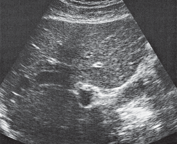
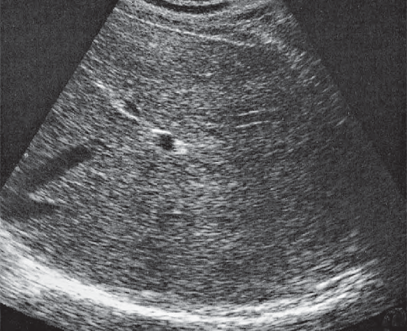
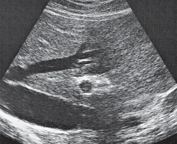
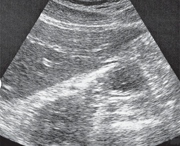
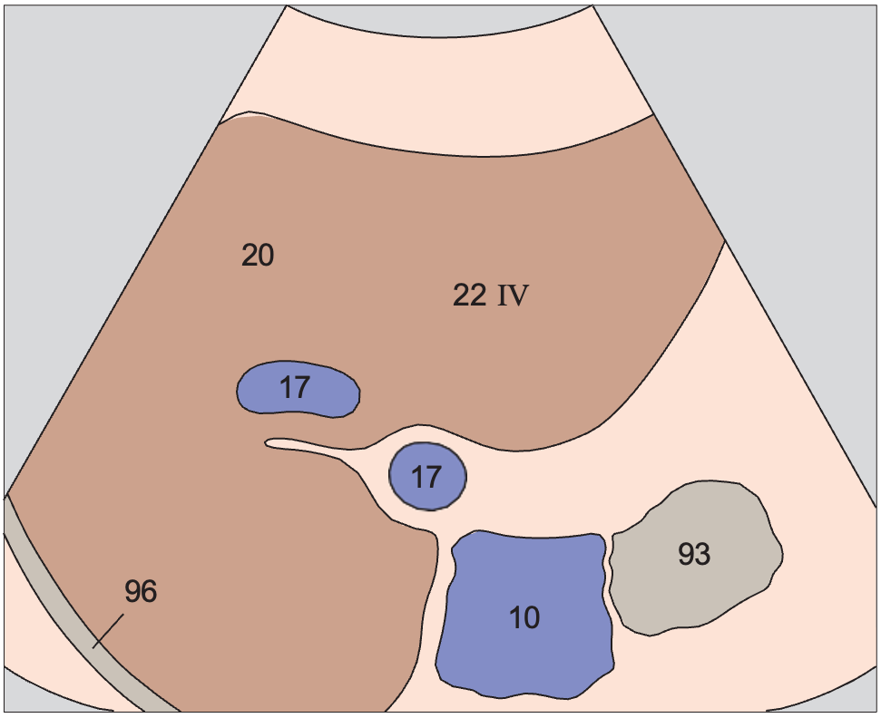
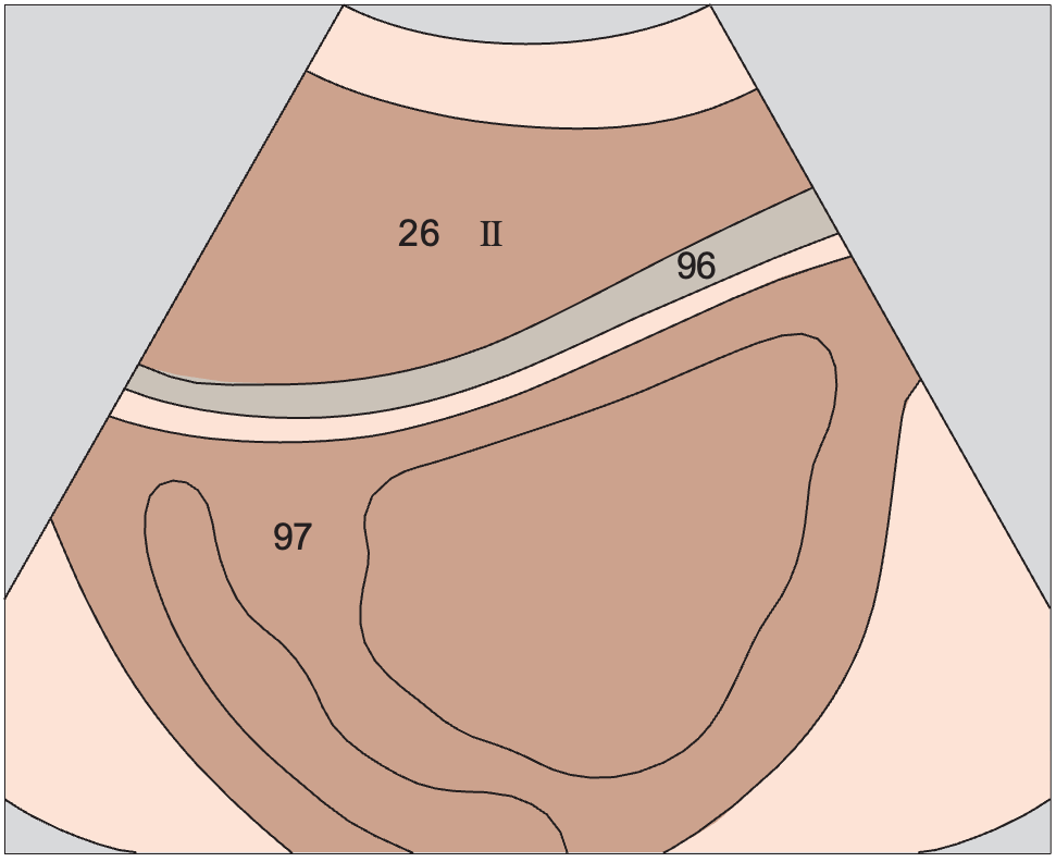
Medial segment ultrasound anatomy is presented here as a labeled cross-sectional atlas. Every view pairs a color-coded anatomy diagram with the matching real ultrasound image, so you can learn the expected shape and position of the medial segment and then recognise it on a real scan.
In the atlas legend the medial segment belongs to the liver group and is shown in a consistent colour wherever it appears. The small body marker on each card shows where the transducer sits for that view, which makes it easier to reproduce the image at the bedside.
Medial segment across the atlas
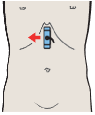
Medial segment, subsegment IV, quadrate lobe
Liver / Liver in Longitudinal Sections · Longitudinal plane
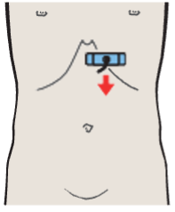
Left lobe of liver, lateral segment, heart
Liver / Left Portions of the Liver in Transverse Sections · Transverse plane
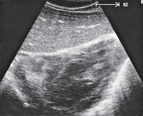
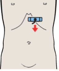
Left lobe of liver, lateral segment, hepatic veins
Liver / Left Portions of the Liver in Transverse Sections · Transverse plane
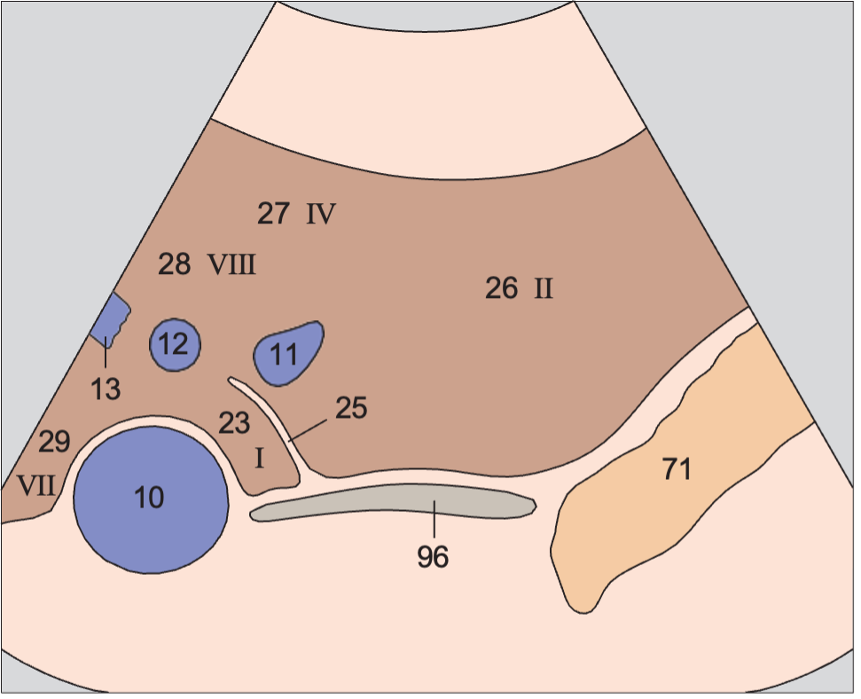
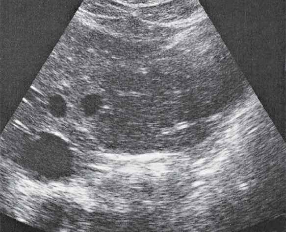
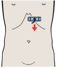
Left lobe of liver, lateral segment, caudate lobe
Liver / Left Portions of the Liver in Transverse Sections · Transverse plane
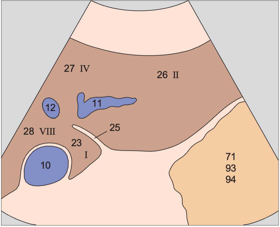
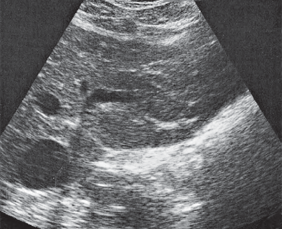
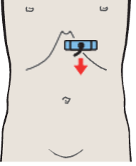
Left lobe of liver, lateral segment, caudate lobe
Liver / Left Portions of the Liver in Transverse Sections · Transverse plane
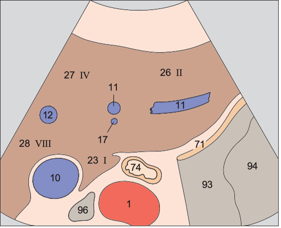
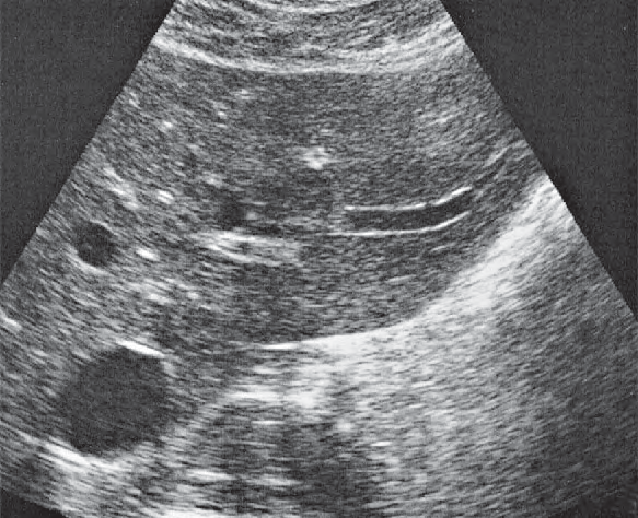
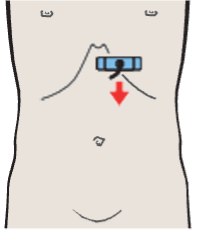
Left lobe of liver, lateral segment, left portal vein branch
Liver / Left Portions of the Liver in Transverse Sections · Transverse plane
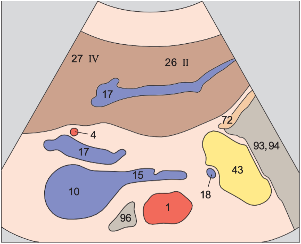
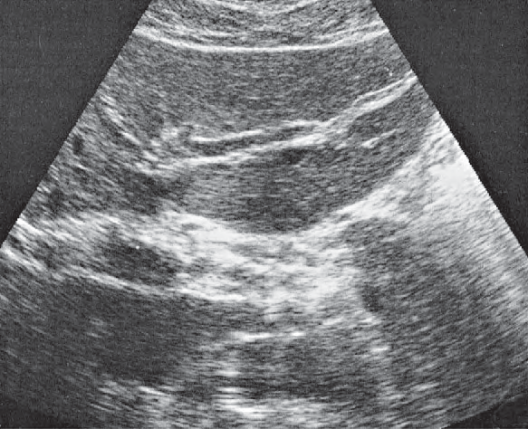
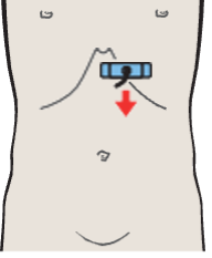
Left lobe of liver, lateral segment, ligamentum teres
Liver / Left Portions of the Liver in Transverse Sections · Transverse plane
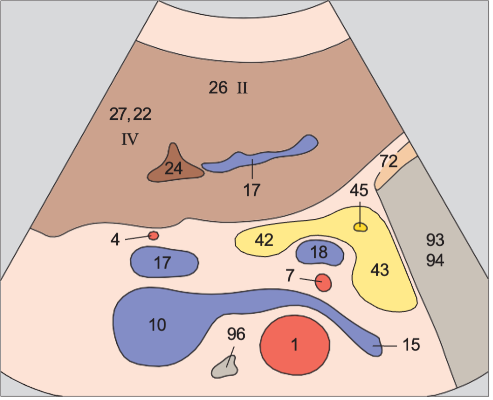
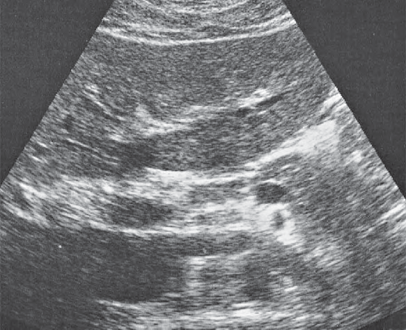
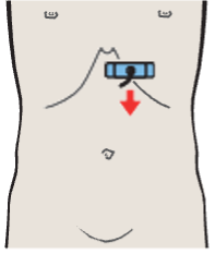
Left lobe of liver, subsegment III, ligamentum teres
Liver / Left Portions of the Liver in Transverse Sections · Transverse plane
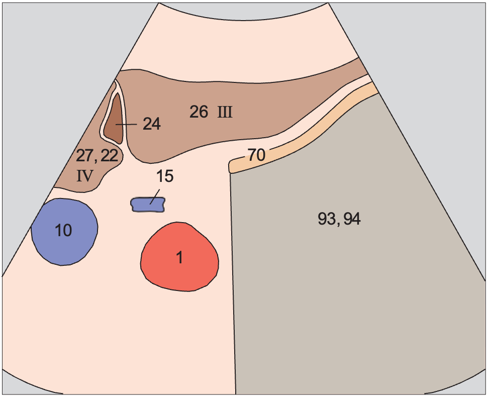
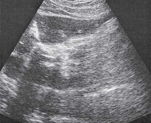
Anatomy diagrams, ultrasound images and probe positions are part of the Ultrasound Anatomy atlas. Colours follow the atlas legend, grouping vessels, organs and other structures by type.